The human eye—a marvel of biological engineering—is also remarkably vulnerable to a wide array of infections. From the common “pink eye” that spreads rapidly among schoolchildren to sight-threatening corneal ulcers and orbital cellulitis, eye infections can range in severity from mild discomfort to true medical emergencies. Early recognition, proper diagnosis, and prompt treatment are essential not only to relieve pain and inflammation but also to preserve your vision. This guide provides an in-depth look at the most prevalent eye infections, explores their causes and clinical presentations, and reviews current best practices for treatment and prevention.
1. Anatomy of the Eye and Routes of Infection
To understand how infections take hold, it helps to recall basic eye anatomy:
-
Conjunctiva: The thin, transparent membrane lining the eyelids and covering the white of the eye.
-
Cornea: The clear, dome-shaped front surface of the eye that focuses light.
-
Eyelids and Eyelashes: Provide protection; contain oil glands (meibomian glands) that maintain tear-film stability.
-
Orbital Tissues: Fat, muscle, and connective tissue that support the eyeball within the bony orbit.
Microbes may invade the eye via:
-
Direct Contact—Touching or rubbing your eyes with contaminated hands.
-
Droplet Spread—Respiratory viruses landing on the conjunctiva (e.g., adenovirus).
-
Contact Lenses—Improper cleaning fosters bacterial or amoebic growth on lens surfaces.
-
Trauma—Scratches or foreign bodies introduce pathogens directly to deeper tissues.
-
Hematogenous Spread—Bloodborne bacteria or fungi seed the internal eye in immunocompromised patients.
2. Bacterial Conjunctivitis (“Pink Eye”)
2.1 Overview
Bacterial conjunctivitis is among the most common eye infections, especially in children. Typical culprits include Staphylococcus aureus, Streptococcus pneumoniae, and Haemophilus influenzae.
2.2 Clinical Features
-
Redness: Diffuse injection of the conjunctival vessels.
-
Purulent Discharge: Thick, yellow-green; matting of eyelids on waking.
-
Foreign-Body Sensation: Gritty feeling.
-
No Significant Pain: Generally mild irritation rather than severe pain.
2.3 Diagnosis
Primarily clinical. In refractory or severe cases, an ophthalmologist may swab the conjunctiva for culture and sensitivity testing.
2.4 Treatment
-
Topical Antibiotics:
-
Erythromycin ointment twice daily for 5–7 days.
-
Fluoroquinolone drops (e.g., moxifloxacin) four times daily in adults.
-
-
Warm Compresses: Apply 5-10 minutes, 3–4 times daily to soften discharge.
-
Hygiene Measures:
-
Frequent handwashing.
-
Dispose of or disinfect pillowcases, towels, and eye makeup.
-
Patients typically improve within 48–72 hours of starting antibiotics. If no improvement occurs, reassessment for resistant bacteria or alternative diagnoses is warranted.
3. Viral Conjunctivitis
3.1 Overview
Adenoviruses cause up to 80% of viral conjunctivitis cases. It often accompanies an upper respiratory infection or cold.
3.2 Clinical Features
-
Watery Discharge: Clear, copious tearing.
-
Preauricular Lymphadenopathy: Swollen lymph node in front of the ear.
-
Hemorrhagic Spots: Subconjunctival hemorrhages in severe epidemic forms.
-
Photophobia and Itching: Mild to moderate discomfort.
3.3 Diagnosis
Primarily based on history and exam. PCR assays of tear fluid can confirm adenovirus but are rarely needed in routine cases.
3.4 Treatment
-
Supportive Care Only:
-
Cool compresses for comfort.
-
Lubricating artificial tears hourly if needed.
-
-
Strict Isolation:
-
Stay home from work/school until discharge stops (usually 1–2 weeks).
-
-
Antiviral Therapy:
-
Rarely indicated except in severe keratoconjunctivitis—topical cidofovir under specialist guidance.
-
Recovery is spontaneous, but vigilant hygiene prevents transmission. Avoid corticosteroid eye drops, which can prolong viral shedding and worsen outcomes.
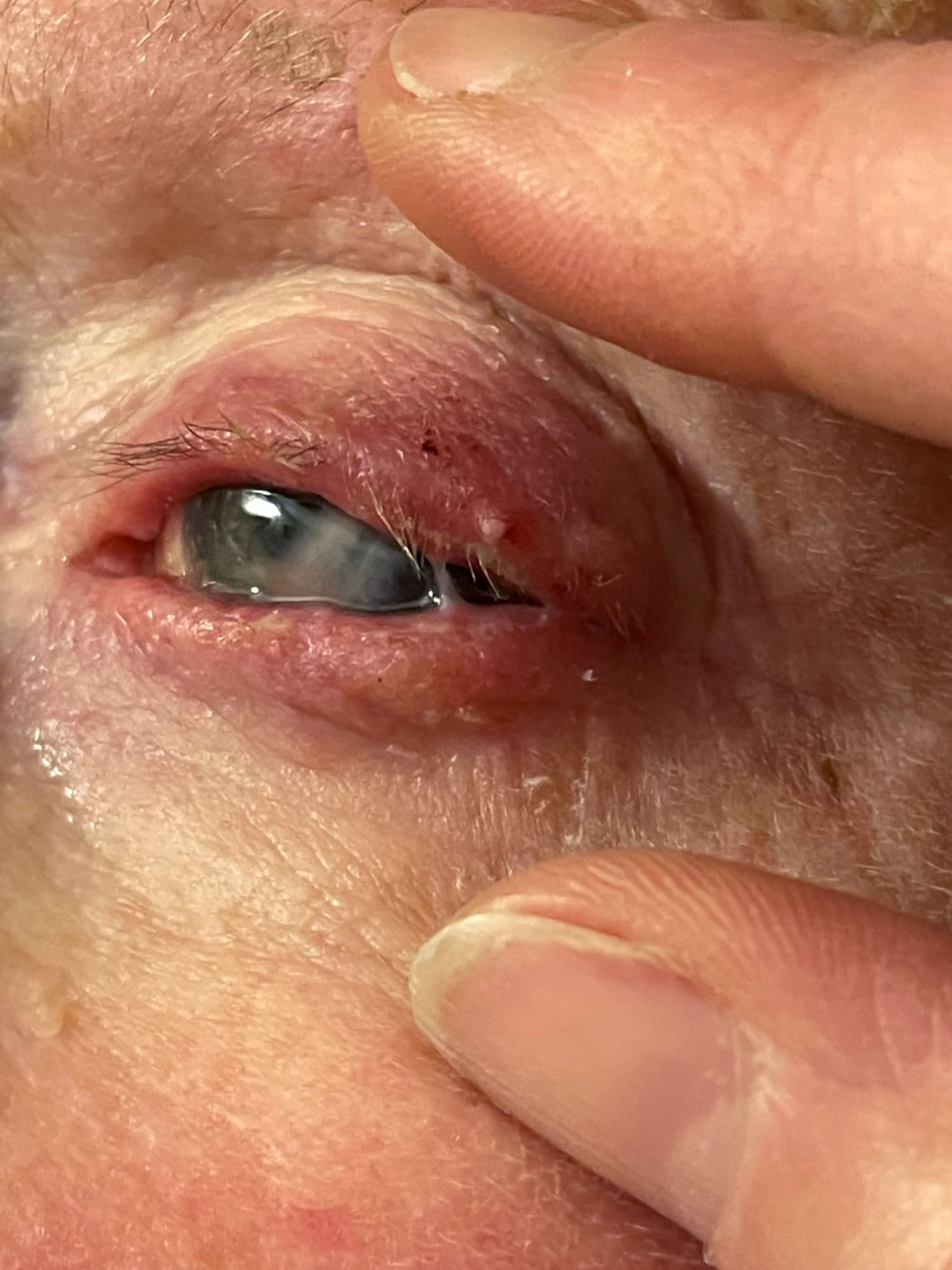
4. Blepharitis and Demodex Infestation
4.1 Blepharitis (Eyelid Margin Inflammation)
Chronic bacterial overgrowth and skin conditions can inflame the eyelid margins.
-
Symptoms: Itching, burning, crusting, and red swollen lid edges.
-
Management:
-
Daily lid hygiene with warm compresses and dilute baby shampoo scrubs.
-
Topical antibiotic ointment (bacitracin or erythromycin) at night.
-
Oral doxycycline (100 mg daily) for 4–6 weeks in refractory or rosacea-associated cases.
-
4.2 Demodex Mite Overgrowth
Tiny mites (Demodex folliculorum and Demodex brevis) live in lash follicles; overgrowth can mimic blepharitis.
-
Signs: Cylindrical dandruff at lash base, intense itching.
-
Treatment: Tea-tree oil-based lid foams or microsponge gels kill mites. Repeat applications weekly for 6–8 weeks.
5. Hordeolum (Stye) and Chalazion
5.1 Hordeolum (Acute Stye)
An acute bacterial infection of an eyelash follicle or meibomian gland.
-
Presentation: Painful, red bump on lid margin; tenderness.
-
Treatment:
-
Warm compresses for 10–15 minutes, 3–5 times daily.
-
Topical antibiotic ointment if secondary infection suspected.
-
Incision and drainage by an ophthalmologist if no resolution in 1–2 weeks.
-
5.2 Chalazion
A chronic, noninfectious blockage of a meibomian gland.
-
Presentation: Painless, firm eyelid nodule away from lash line.
-
Treatment:
-
Warm compresses and lid massage.
-
Intralesional corticosteroid injection if persistent.
-
Surgical excision under local anesthesia for large or refractory chalazia.
-
6. Keratitis (Corneal Infection)
6.1 Bacterial Keratitis
Often linked to contact-lens misuse. Rapidly progressive and potentially blinding.
-
Symptoms: Severe pain, redness, discharge, blurred vision, light sensitivity.
-
Management:
-
Urgent referral to ophthalmology.
-
Intensive topical antibiotics (hourly fortified cephalosporin + aminoglycoside).
-
Avoid steroids until infection controlled.
-
6.2 Viral Keratitis
Herpes simplex virus invades the cornea, causing dendritic ulcers.
-
Signs: Branching corneal ulcer on fluorescein stain.
-
Treatment:
-
Topical or oral antivirals (trifluridine drops, oral acyclovir).
-
Close monitoring to prevent corneal scarring.
-
6.3 Fungal Keratitis
Tropical climates and plant trauma predispose to Fusarium or Aspergillus infections.
-
Symptoms: Feathery-margined ulcer, satellite lesions.
-
Treatment:
-
Natamycin 5% drops hourly for filamentous fungi.
-
Voriconazole drops for yeasts.
-
Possible corneal debridement to enhance drug penetration.
-
6.4 Acanthamoeba Keratitis
Associated with poor contact-lens hygiene, particularly homemade saline.
-
Presentation: Severe, disproportionate pain, ring infiltrate on cornea.
-
Treatment:
-
Biguanide (polyhexamethylene biguanide) 0.02% and diamidine (propamidine) drops.
-
Treatment duration: 3–6 months under specialist care.
-
7. Endophthalmitis and Orbital Cellulitis
7.1 Endophthalmitis
A rare but catastrophic infection of the eye’s interior, often post-surgical or traumatic.
-
Symptoms: Sudden vision loss, pain, severe inflammation inside the eye.
-
Management:
-
Emergency intravitreal antibiotics.
-
Vitrectomy may be required to clear infection.
-
7.2 Orbital Cellulitis
Infection of the tissues behind the orbital septum—an emergency requiring hospitalization.
-
Signs: Eyelid swelling, pain with eye movement, fever, possible vision impairment.
-
Treatment:
-
Intravenous broad-spectrum antibiotics (vancomycin + ceftriaxone).
-
CT or MRI to assess for abscess.
-
Surgical drainage of any abscess.
-
8. Diagnosis: When to See a Specialist
-
Mild Conjunctivitis: Often managed by primary care or optometrist.
-
Any Keratitis: Immediate ophthalmology referral—risk of corneal scarring.
-
Orbital Cellulitis: Emergency department.
-
Endophthalmitis: Ophthalmology/emergency department without delay.
Diagnostic tools include slit-lamp examination, fluorescein staining of the cornea, conjunctival swabs, and imaging (CT/MRI) for orbital infections.
9. Adjunctive Measures and Home Care
-
Warm and Cold Compresses: Soothe inflammation, improve comfort.
-
Lubricating Eye Drops: Preserve tear-film integrity.
-
Strict Lid Hygiene: Daily cleaning in blepharitis.
-
Proper Contact-Lens Hygiene:
-
Wash hands before handling lenses.
-
Use only sterile solutions; never tap-water.
-
Discard lenses and cases per manufacturer guidelines.
-
-
Avoid Eye Rubbing: Transfers bacteria and viruses.
10. Prevention Strategies
-
Hand Hygiene: The cornerstone of preventing conjunctivitis.
-
Avoid Sharing Personal Items: Towels, cosmetics, contact-lens cases.
-
Protective Eyewear: Safety goggles in dusty or chemical environments.
-
Vaccinations: MMR vaccine reduces risk of measles-related ocular complications.
-
Regular Eye Exams: Early detection of dry eye, blepharitis, or other predisposing conditions.
11. Emerging Therapies and Research
-
Antimicrobial Peptides: New topical agents in development for drug-resistant bacteria.
-
Phage Therapy: Bacteriophages targeting Pseudomonas in keratitis trials.
-
Drug-Eluting Contact Lenses: Microdose delivery of antibiotics directly to the cornea.
-
Immunomodulatory Agents: For herpes stromal keratitis to reduce scarring.
12. Conclusion
Eye infections pose a multifaceted threat to ocular health. While many cases—such as mild bacterial or viral conjunctivitis—resolve with basic hygiene and antibiotic or supportive care, others (keratitis, endophthalmitis, orbital cellulitis) demand urgent specialist intervention. Understanding the spectrum of eye infections, recognizing early warning signs, and implementing prompt, evidence-based treatments are essential to preserving vision and preventing complications. Embrace preventive measures—rigorous hand hygiene, safe contact-lens practices, and protective eyewear—and maintain regular eye examinations to support long-term ocular well-being. When in doubt, consult an ophthalmologist: rapid diagnosis and targeted therapy remain the best defenses against sight-threatening infections.